Acoustic Neuroma What is an Acoustic Neuroma? How is the tumor identified? What treatments are available for acoustic
neuroma?
Observation
Surgical Treatment
Stereotactic Radiation
What is an Acoustic Neuroma?
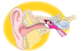
An acoustic neuroma (also called a schwannoma or neurinoma) is a benign (non-cancerous)
tissue growth that arises from the balance nerve. There are two balance (vestibular)
nerves, one for each of the right and left ears. The balance nerve is a thin
cord that starts at the base of the brain and travels through a small, bony
canal (internal auditory canal) of the skull to the inner ear. Two other
nerves, the hearing nerve and the facial nerve (7th cranial nerve), travel
directly alongside the balance nerve through the internal auditory canal
to the inner ear.
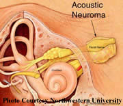 |
| Small acoustic neuroma in the internal auditory canal. |
Acoustic neuromas commonly develop inside the internal auditory canal.
These tumors usually grow slowly over a period of many years.
When acoustic neuromas grow in size, the tumor compresses the
hearing nerve. Therefore, hearing loss is the first symptom in
over 90% of patients with acoustic neuroma. Tinnitus or ear noise
is also a common symptom. Loss of balance, dizziness or facial
weakness may develop as the tumor continues to grow.
With appropriate tests it is now possible to identify tumors as small
as 1-3mm in size. The first step in identifying a possible tumor
is a thorough history and physical examination by a physician. Hearing
testing is performed to identify any loss of hearing or speech understanding.
An auditory brainstem response test (abbreviated ABR, BAER, or BSER)
is ordered if there are any asymmetries in hearing testing between
the right and left ears. An ABR provides information on the passage
of an electrical impulse along the hearing nerve from the ear to
the brain. Abnormal ABR results suggest a poorly functioning hearing
nerve. A detailed "imaging" is ordered if there is an abnormality
of the ABR test.
The most accurate imaging technique for acoustic neuroma identification
is a magnetic resonance imaging (MRI). A properly performed MRI
can identify acoustic neuromas as small as 2-3mm. MRI uses magnetic
pulses and radio frequency waves to produce an image of a portion
of the body being studied. Radiation is not used to perform an
MRI. A contrast material, gadolinium, is given during the MRI
to enhance the visibility of the tumor.
Observation
Since acoustic neuromas are benign, often slow-growing tumors, careful
observation over a period of time may be appropriate for some patients.
When a small tumor is discovered in an older patient, observation to study
the growth rate of the tumor may be indicated if acute symptoms are not
present. A MRI scan of the tumor and surrounding region must be performed
periodically to determine if there is any significant change in the
size of the tumor. If the tumor does not grow, observation is continued.
If the tumor increases in size, treatment may be recommended.
Surgical Removal
There are several surgical approaches that may be used to remove an acoustic
tumor. The type of approach is individualized depending on the patient's
wishes, hearing level, other neurological symptoms, and location and size
of the tumor. The Ohio Ear Institute, LLC is well versed in all surgical
approaches for acoustic neuroma management.
Middle Fossa Approach
In the middle fossa approach, the bone is opened above the ear and the bone
overlying the tumor is removed. The inner ear is not entered. Therefore,
hearing preservation is possible. The middle fossa approach is most suitable
for small tumors with good hearing.
Retrosigmoid Approach
In this approach, the bone is opened behind both the mastoid bone and the
inner ear. In this way the tumor is removed from behind the inner ear. Retrosigmoid
removal also allows the possibility of hearing preservation and may be used
for both small and large tumors.
Translabyrinthine Approach
In the translabyrinthine approach, the mastoid bone behind the ear is removed
and the inner ear is opened. This exposes the internal auditory canal directly.
All hearing is lost with this approach. The translabyrinthine approach is,
therefore, used only for those cases where hearing loss is already severe
or the tumor is so large that hearing conservation is not a realistic goal.
Hospital stay after any type of microsurgery ranges from 4-7 days. Approximately
4-6 weeks is suggested for recovery.
back to the top
Stereotactic Radiation Therapy
Stereotactic radiation therapy, which is commonly known as "radiosurgery," is
a single session radiation treatment. Using computer imaging, a single,
high dose of radiation is delivered to the acoustic tumor while minimizing
injury to surrounding nerves and brain tissue. Treatment is often
performed on an outpatient basis.
Stereotactic radiation prevents further tumor growth and sometimes shrinks
a portion of the tumor. This type of treatment does not remove the tumor.
Follow-up imaging studies are important because some tumors will continue
to grow after stereotactic radiation. For more information regarding Stereotactic Radiation Therapy, please click here.
Each form of acoustic neuroma treatment, either surgery or radiation, has
advantages and disadvantages. Many factors are used to determine the most
appropriate type of treatment.
|