|
|
Gamma Knife Radiosurgery Steps of the Gamma Knife Radiosurgery Procedure Stereotactic Radiosurgery using the Gamma Knife consists of five main steps.
- Frame placement. A lightweight frame is fitted over the patient’s head and attached with screws using local anesthesia. The frame serves two purposes: On the side of the frame are markers that allow the treatment team to pinpoint areas that will receive treatment. The frame also keeps the patient’s head immobile during the radiation procedure.
|
Head frame with CT/MRI box |
- Imaging Study. Imaging studies are obtained using MRI, CT scan, angiography, or a combination of these technologies. The imaging studies produce a three-dimensional map of the tumor and the surrounding brain structures.
|
|
3-D image of a 3-cm acoustic neuroma
(top view)
|
3-D image of a 3-cm acoustic neuroma (frontal view) |
- Treatment planning. Based on the imaging studies, your physician works with a radiation oncologist and a medical physicist to develop the treatment plan that accurately targets the tumor with exact precision, sparing surrounding tissue as much as possible (i.e. a plan than conforms closely to the shape of the lesion).
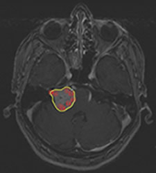 |
Treatment plan for 3-cm acoustic neuroma. Yellow=outline of tumor; red circles=planned radiation shots
|
- Radiation treatment. Treatment begins by first having the patient lie on the Gamma Knife bed (Left Figure Below). The head frame is fitted into a helmet with 201 holes in the helmet (Right Figure Below). The helmet helps to guide 201 sources of cobalt 60 based (photon) radiation beams to accurately target the tumor. After the helmet is placed, the bed is precisely moved into the domed unit and treatment begins. Depending on the size and location of the tumor, the actual time of radiation delivery could range from a few minutes to over an hour.
 |
 |
Leksell 4C gamma knife unit |
Patient insided helmet |
- Removal of the head frame. Removal of the frame is a painless procedure. Most patients are able to go home shortly after the head frame is removed.
|